Wednesday, January 27, 2016

Unit 5 Reflection

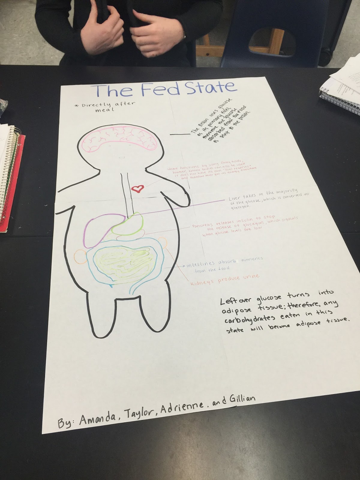
Fed State Poster
In this unit we learned about the digestive system, metabolism, diabetes, endocrine system and the lymphatic system.  We learned that there are six basic functions of digestion, ingestion, secretion, mixing/propulsion, digesting, absorption, defecation. The digestive system has many accessory organs and we found out the pathway that food takes. ATP is the universal carrier for metabolism and it can be generated by the oxidation of glucose, fatty acids and amino acids. It takes three stages for energy to be extracted from food. We learned more about diabetes and what causes it.  Diabetes is when your body cant properly control blood glucose levels and you body cant make enough insulin, and it cant respond to it. Type 1 diabetes is when the body doesn't produce insulin at all and they must take insulin by injecting it. Type 2 diabetes begins with insulin resistance and is more common then type 1 diabetes.  The endocrine system controls the processes involved in movement and physiological equilibrium.  We learned about the organs in the endocrine system and their hormones. We were taught about the lymphatic system and how it is involved in immunity, lipid absorption and fluid recovery.  Lymph is a clear colorless fluid that flows throughout lymphatic vessels.  The main organs of the lymphatic system are Lymph nodes, thymus, tonsils and spleen. In this unit we did a lab where we measured out our digestive tracts with strings, and we found out that it is a lot longer then we thought it was.
strings across the top are digestive tracts

Wednesday, January 6, 2016

The Digestion System Lab

In this lab we measured out the length of each part of the digestive system and at the end we added up all of the lengths and got the length of our digestive tracts. The main takeaways I got from this activity is that the intestines are a lot longer than I imagined.  With all of the digestive lengths put together it was very long.  For our small intestine we multiplied our height times 4, which is pretty long so for it to be able to fit in our abdomen it needs to be very bunched up and folded.  The small intestine is very long but it is not as wide as the large intestine.  The large intestine is much wider than the small intestine but it is shorter, for this your large intestine was about your height so we used out height and measured out the yarn.  My guess to how long it takes for food to move through your entire digestive system would be about 8 hours.  I looked it up and it says it takes between 24-72 hours for food to move through the entire system.  My guess was very off, but a big factor to how long it takes is the type of food it is.  Digestion is different from absorption because digestion is breaking down food into smaller pieces and absorption is taking the needed nutrients. Your intestines are where absorption takes place and digestion involves the mouth, esophagus, stomach, small intestine, large intestine, liver, gall bladder, pancreas, anal cavity and the anus.  I don't have any questions about digestion, I want to learn more about how different people can't digest foods, but others can like for example celiac disease.

Monday, January 4, 2016

New Years Goals

My first goal for the new year is I need to start putting more time aside to study.  I need to be more on track with when my tests and quizzes are and plan ahead to make sure that I have enough time to go over everything and have time to study well. I also need to study more thoroughly for all of my classes. My second goal is I need to get more sleep.  For me to get more sleep I need to go to bed earlier, which means I have to get my homework done earlier so I am not up late trying to finish it.  I usually procrastinate when I get home from school, but I want to start getting my homework done quicker so I have more time so I can go to bed earlier.  It will also help me be more awake during school.

Thursday, November 12, 2015

Sheep Heart Dissection Reflection Questions

1. What is the purpose of the pericardium?
The pericardium is a sac of serous membranes that secretes a fluid to lubricate the heart and reduce friction.  It is made of visceral and parietal layers.
2. How do arteries differ from veins in their structure? 
Veins carry blood from the tissues to the heart and arteries carry blood away from the heart to the tissues.  Veins have thinner walls, arteries are larger and are under higher pressure.
3. What function does the auricles serve?
They are chambers that blood enters the heart.
4. What external differences of the atria and ventricles do you observe? 
The walls of the ventricles are thicker, the atrium's are above the ventricles and they are smaller than the ventricles.
5. Find the following structures, and use pictures or words to describe each.
a. Coronary Sinus: receives blood from the coronary veins, and flows into the right atrium.
b. Inferior Vena Cava: Large vein that carries deoxygenated blood into the heart. Carries blood from the lower body.
c. Right Atrioventricular Valve (Tricuspid Valve): It allows blood to flow in one direction and it prevents back flow of blood. The Tricuspid Valve is between the right atrium and the right ventricle.
6. Draw a picture of the Tricuspid Valve including the chordate tendinae and the papillary muscle.

7. Why is the "anchoring" of the heart valves by the chordate tendineae and the papillary muscle important to heart function? They are there to prevent the tricuspid and bicuspid valves from collapsing into the atrium when it contracts.
8. Using pictures and/or words, describe what you see (bicuspid valve): It prevents back flow of blood, and allows blood to flow in one direction, it is between the left atrium and the left ventricle, and is also called the mitral valve.
9. What is the function of the semi-lunar valves? Semi-lunar valves prevents arterial blood from re-entering the heart.
10.  a. If the valve disease occurs on the right side of the heart, it results in swelling in feet and ankles, why might this happen? Because the right side of the heart deals with deoxygenated blood, so the feet and ankles are not getting oxygenated blood.
b. If valve disease occurs on the left side of the heart, what complications would you expect to see?
The left side of the heart deals with oxygenated blood, so deoxygenated blood would be filling up the lungs, so the lungs would swell.
11. Using pictures or words, describe what you see: The coronary tendineae connects the papillary muscle to the bicuspid valve.
12. Describe the left and right sides of the heart differ from each other? 
The right side of the heart deals with deoxygenated blood and the left side of the heart deals with oxygenated blood.  The right side of the heart goes to the lungs and the left side comes from the lungs.
13. Draw and label all structures visible in the interior of the cross section.


Wednesday, November 11, 2015

Unit 3 Reflection

This unit taught us all about the circulatory system, the major components, specific parts, accessory organs, and much more.  The cardiovascular system is a  fluid filled network of tubes or vessels, through which materials move between the environment and the cells of a multicellular animal.  Cardiovascular health is making sure that you keep your heart healthy.  Theres several things that cause heart attacks, a heart attack is when blood supply to the myocardium is severely reduced, or stopped resulting in damage to the heart cells or death of heart cells. This is caused by one or more coronary arteries blocked, usually due to atherosclerosis.  Stroke is a brain injury that occurs when blood supply to part of the brain is interrupted. This is also often caused by atherosclerosis, but can be caused by an aneurysm.  To promote your cardiovascular health, you can control your blood pressure, stop smoking, control cholesterol, eat a healthy diet, drink alcohol in moderation, control diabetes, control your weight, exercise, manage your stress, and your genetic history.  I think this unit was very interesting because it applies to our lives, and it helped me understand what happens when these things go wrong.  Also it taught me ways of identifying these situations, which is important to know.  This unit we did our first dissection, and it was helpful to actually see the parts of the heart it made it easier to understand how it works.  For the remainder of the semester, I want to study more and the temp checks are very helpful, so its nice when we have them because it lets me know how much information I retained, so my goal is to study more and do better on tests.
Sheep heart dissection
Heart Diagram 

Atherosclerosis

Monday, October 26, 2015

Monday Wellness: Snacking

The snack we made during class was healthy because we used apples which are a good source of vitamin c,and organic peanut butter which has no trans fats, and it almonds which the peanut butter and almonds has a lot of iron and magnesium.  I usually eat snacks every couple of hours, depending on when i'm hungry. A healthy snack is something that isn't artificially flavored.  Snacks to stay away from would probably things with a lot of empty calories and with little nutritional values.

Tuesday, October 13, 2015

Blood Pressure Lab

Systolic Blood pressure is the first number you record when taking someones blood pressure. For example if your blood pressure is 120/80 the systolic blood pressure is 120.  It represents the pressure when the heart contracts.  Your diastolic blood pressure is the second number and it is when the heart relaxes.  We used a stethoscope to measure our heart rates, and to measure our blood pressure we used stethoscope and a sphygomomanometer. Using your thumb to measure blood pressure is not ideal because your thumb has a heart beat also, so it would interfere with the other heart beat your measuring. To use a sphygomomanometer you put it on the person's upper arm whose blood pressure is being measured and you tighten it so its tight but not restricting.  To record the blood pressure you need a stethoscope so you would place it on their inner elbow and the hearing parts in your ears to listen, then you would have one hand on the stethoscope and your other hand holding the bulb that you squeeze your hand and it pumps air into the arm cuff. You pump the bulb until its around 180 and then you slowly release the air, you listen for the heart beat and as soon as you hear it you record the first number and then when the heart beat noise stops you record that number too. You put the first number you recorded over the second number you recorded and thats your blood pressure.
Charts 
Blood Pressure Cuff and Stethoscope