In this article it talks about a woman who was unaware of the fact that she is missing her cerebellum until she was 24. She went to the hospital because she was nauseous and dizzy and they did a CAT scan and it showed that where her cerebellum should be it was filled with cerebrospinal fluid. She had trouble walking most of her life and she also wasn't able to speak that well until she was 7. The cerebellum is 10% of the brains volume but 50% of the brains neurons. The cerebellum's function is it controls your voluntary movements and balance.
The Parietal Lobe
The parietal lobe controls sensations, it deals with and reacts to environment and it processes sensory information. If your parietal lobe is damaged, the person will have trouble reading, recognizing things and people. It will be harder for them to be aware of their bodies. People with damaged parietal lobes have trouble multitasking, issues with telling right and left apart and the mathematic skills they have is often reduced. You can't live without the parietal lobe.
Thursday, April 14, 2016

The Clay Brain
We studied the brain and made a model of the left hemisphere along the sagittal plane and the right cerebral hemisphere using clay on cardboard. We traced an outline of the brain with pencil and filled in the spots with different colored clay, then labeled the parts in a key.


Thursday, March 24, 2016
Unit 7 Reflection
 In this unit, we learned about the muscular system. There are a lot of different joints and defining characteristics for them. For example, there are levers, which is a rigid object that rotates around a fixed point called a fulcrum. Rotation occurs when effort overcomes resistance. There are first class levers, second class levers and third class levers. Joints are the point of a connection between two bones or elements of a skeleton especially if it allows motion. Ligaments are bone to bone, and tendons are bone to muscle. The functional classification of joints are synarthroses, or immovable joints, amphiarthroses, or slightly movable joints, and diarthroses, or freely movable joints. There are fibrous joints which are generally immovable, cartilaginous joints, which are immovable or slightly movable, and synovial joints which are freely movable. There are 17 different directional terms and joint movements that we learned about. For example, flexion is the movement that decreases the angle between two bones and extension is the movement that increases the angle between two bones. Muscles are very important in our bodies because they do a lot of things, like the movement of bones, maintaining posture and body position, stabilizing joints, and heat generation. There are a few properties of muscle tissue, excitability, or the ability to receive and respond to stimuli, contractility, or the ability to shorten when stimulated, extensibility, or the ability to be stretched, and elasticity, or the ability to recoil to resting length. Connective tissues components are fascia (thin sheets of fibrous connective tissue that holds muscle fibers together), epimysium (fascia that surrounds the entire muscle), perimysium (separates muscles into bundles), endomysium (fascia that surrounds each muscle fiber), and tendon (a cord of dense regular connective tissue composed of collagen that attaches muscle to periosteum to bone). Origin is the immovable end of the muscle and insertion is the movable end of the muscle, when a muscle contracts the insertion is moved towards the origin. We learned about skeletal muscle, and that its composed of thousands of bundles of muscle fibers. We did a chicken dissection, where we located all of the main muscles and labeled them. We wrote reflections on our blogs. My Chicken Dissection Reflection. Mr. Orre taught us about the three different types of muscle fibers. There slow twitch fibers which are slow oxidative, fast twitch fibers which are fast oxidative, and fast twitch fibers which are fast glycolytic. The general effects of exercise on muscles is it helps to increase metabolism, strengthens bones, increases muscular endurance, helps prevent injuries, and it increases coordination and balance. Hypertrophy is when cells increase in volume due to move myofibrils, and hyperplasia is when cells remain the same in size, but they increase in number. We made a stop motion video to show how muscle contractions work. Our Muscle Contraction Video To keep our muscles healthy it is important to exercise regularly, eat healthy, don't smoke, know basic first aid, and you should train aerobically and anaerobically. We also were taught about performance enhancement substances and how it effects our bodies. This unit, we learned a lot about the muscular system, and it helped me understand it a lot more.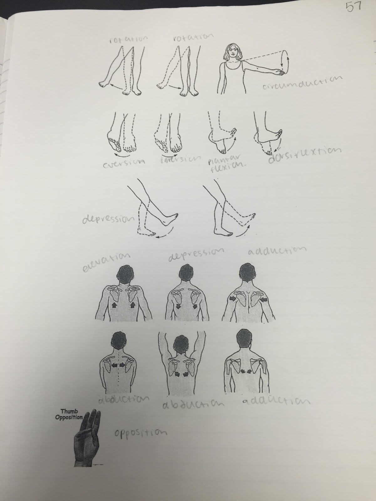
 |
| Picture from our chicken dissection |
Wednesday, March 23, 2016
Human Growth Hormone

Sunday, March 20, 2016
Chicken Disection Analysis

 In this Lab we dissected a chicken and we will be locating the main muscles. We began by removing the skin from the chest. We found the pectoralis major and the pectoralis minor which is right under it. Next we removed the skin on its back and found the trapezius and latissimus dorsi. Next we Removed the skin on the chickens wings and found the deltoid, biceps brachii and the triceps humeralis. We also found the flexor carpi ulnaris and the brachiordialis. Next we removed the skin on the chickens thigh and found the sartorius, iliotibialis, semimembranosus, semitendinous, quadriceps, and the gastrocnemius. We also found the preens longs and the tibias anterior. The pectoralis major helps pull the wing ventrally and the pectoralis minor lifts the wing dorsally. The trapezius helps shrug the shoulders and the latissimus dorsi extends or pulls the wing. The deltoid helps raise the upper wing, the biceps brachii helps bend the wing, and the triceps humeralis extends the wing. The flexor carpi unlearns flexes the hand, and the brachiordialis helps pull the hand back. The sartorius flexes the thigh, the iliotibialis extends the thigh, the biceps femurs flexes the leg and the semimembranosus extends the thigh also. Semitendinosis extends the thigh, quadriceps femurs extends the lower left, and the gastrocnemius is used for standing on your toes. The preens longs extends the foot and the tibias anterior flexes the foot. The biceps brachii is an example of an origin, and origins are the attachment site of the muscles tendon to a stationary bone. Insertion is the attachment site of a tendon to a moveable bone. There are a lot of muscles in the chicken that are pretty similar to the muscles we have. For example the deltoid what is on the center top of the shoulder of the chicken and of humans which helps to raise your upper arm or in chickens their wing. The trapezius is muscles that run perpendicular from the backbone to the shoulder of the bird and pull the shoulder back, but in humans they're split into two parts. The triceps humeralis is the muscle on the inferior side of the upper wing or arm in birds and humans, and it extends the arm or in chickens the wing.


 |
| Add caption |


Monday, February 29, 2016
Unit 6 Reflection
 |
| Owl Pellet lab |
 |
| Skeleton |

 |
| Bone slide |
Thursday, February 25, 2016
Owl Pellet Lab
 |
 |
| humerus |
 We did an Owl Pellet lab, where we dissected owl pellets and tried to figure out what type of animal the owl ate. We had to look at the bones we found and try to match them to other animal bones. Our owl pellet was 5.69 grams, 9 cm long and 2 cm wide. In it we found two skulls and a lot of very small bones. The skulls were 3.5 cm long, 2.5 cm wide and the lower jaw was 2.5 cm long and 1 cm wide. Looking at the charts and information we were given, we decided that it was a vole. The first thing we saw was the humerus, the bone has a part that pokes out and it matched the picture we had of a vole's humerus. Then the skull has a curve right after the front of the mouth and then in the back there were teeth and the vole was the only animal that has a skull with this shape. Also the lower jaw we found has the same shape as a vole. The ribs that we found in our owl pellet are very similar to ribs on a human, they are the same shape. The back bones we found were similar to human back bones because they lined up like human spines do. We found a bone which we thought was the femur and it was pretty similar to the human femur, it was the same shape and had the ball in socket joint. Some things we found different was the humerus, the bone poked out at a point and the human bone doesn't do that. Also the tibia and fibula were reversed, in the human skeleton the tibia is bigger than the fibula but in the vole, the fibula is bigger than the tibia and they were also connected. Another difference we found was in the skull, it has very big eye sockets and the mouth is a lot different. Overall we learned a lot about the differences in these bones and human bones, and also it was interesting to see what owls eat and to take apart the owl pellets.
 |
| tibia and fibula |

 |
| skull and mandible |
Subscribe to:
Posts (Atom)