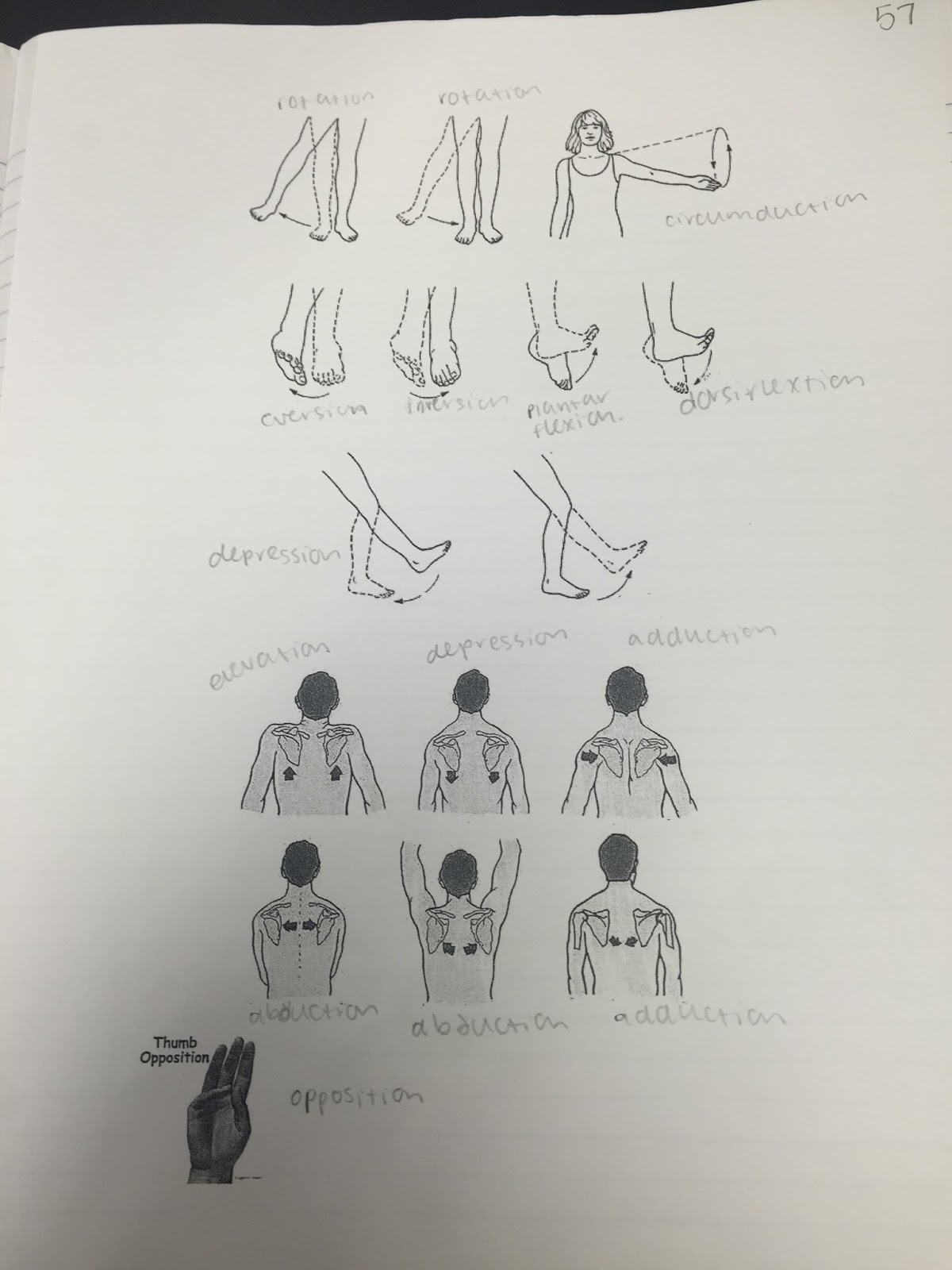
In this unit, we learned about the muscular system. There are a lot of different joints and defining characteristics for them. For example, there are levers, which is a rigid object that rotates around a fixed point called a fulcrum. Rotation occurs when effort overcomes resistance. There are first class levers, second class levers and third class levers. Joints are the point of a connection between two bones or elements of a skeleton especially if it allows motion. Ligaments are bone to bone, and tendons are bone to muscle. The functional classification of joints are synarthroses, or immovable joints, amphiarthroses, or slightly movable joints, and diarthroses, or freely movable joints. There are fibrous joints which are generally immovable, cartilaginous joints, which are immovable or slightly movable, and synovial joints which are freely movable. There are 17 different directional terms and joint movements that we learned about. For example, flexion is the movement that decreases the angle between two bones and extension is the movement that increases the angle between two bones. Muscles are very important in our bodies because they do a lot of things, like the movement of bones, maintaining posture and body position, stabilizing joints, and heat generation. There are a few properties of muscle tissue, excitability, or the ability to receive and respond to stimuli, contractility, or the ability to shorten when stimulated, extensibility, or the ability to be stretched, and elasticity, or the ability to recoil to resting length. Connective tissues components are fascia (thin sheets of fibrous connective tissue that holds muscle fibers together), epimysium (fascia that surrounds the entire muscle), perimysium (separates muscles into bundles), endomysium (fascia that surrounds each muscle fiber), and tendon (a cord of dense regular connective tissue composed of collagen that attaches muscle to periosteum to bone). Origin is the immovable end of the muscle and insertion is the movable end of the muscle, when a muscle contracts the insertion is moved towards the origin. We learned about skeletal muscle, and that its composed of thousands of bundles of muscle fibers. We did a chicken dissection, where we located all of the main muscles and labeled them. We wrote reflections on our blogs.
My Chicken Dissection Reflection. Mr. Orre taught us about the three different types of muscle fibers. There slow twitch fibers which are slow oxidative, fast twitch fibers which are fast oxidative, and fast twitch fibers which are fast glycolytic. The general effects of exercise on muscles is it helps to increase metabolism, strengthens bones, increases muscular endurance, helps prevent injuries, and it increases coordination and balance. Hypertrophy is when cells increase in volume due to move myofibrils, and hyperplasia is when cells remain the same in size, but they increase in number. We made a stop motion video to show how muscle contractions work.
Our Muscle Contraction Video To keep our muscles healthy it is important to exercise regularly, eat healthy, don't smoke, know basic first aid, and you should train aerobically and anaerobically. We also were taught about performance enhancement substances and how it effects our bodies. This unit, we learned a lot about the muscular system, and it helped me understand it a lot more.

 |
| Picture from our chicken dissection |
 In this unit, we learned about the muscular system. There are a lot of different joints and defining characteristics for them. For example, there are levers, which is a rigid object that rotates around a fixed point called a fulcrum. Rotation occurs when effort overcomes resistance. There are first class levers, second class levers and third class levers. Joints are the point of a connection between two bones or elements of a skeleton especially if it allows motion. Ligaments are bone to bone, and tendons are bone to muscle. The functional classification of joints are synarthroses, or immovable joints, amphiarthroses, or slightly movable joints, and diarthroses, or freely movable joints. There are fibrous joints which are generally immovable, cartilaginous joints, which are immovable or slightly movable, and synovial joints which are freely movable. There are 17 different directional terms and joint movements that we learned about. For example, flexion is the movement that decreases the angle between two bones and extension is the movement that increases the angle between two bones. Muscles are very important in our bodies because they do a lot of things, like the movement of bones, maintaining posture and body position, stabilizing joints, and heat generation. There are a few properties of muscle tissue, excitability, or the ability to receive and respond to stimuli, contractility, or the ability to shorten when stimulated, extensibility, or the ability to be stretched, and elasticity, or the ability to recoil to resting length. Connective tissues components are fascia (thin sheets of fibrous connective tissue that holds muscle fibers together), epimysium (fascia that surrounds the entire muscle), perimysium (separates muscles into bundles), endomysium (fascia that surrounds each muscle fiber), and tendon (a cord of dense regular connective tissue composed of collagen that attaches muscle to periosteum to bone). Origin is the immovable end of the muscle and insertion is the movable end of the muscle, when a muscle contracts the insertion is moved towards the origin. We learned about skeletal muscle, and that its composed of thousands of bundles of muscle fibers. We did a chicken dissection, where we located all of the main muscles and labeled them. We wrote reflections on our blogs. My Chicken Dissection Reflection. Mr. Orre taught us about the three different types of muscle fibers. There slow twitch fibers which are slow oxidative, fast twitch fibers which are fast oxidative, and fast twitch fibers which are fast glycolytic. The general effects of exercise on muscles is it helps to increase metabolism, strengthens bones, increases muscular endurance, helps prevent injuries, and it increases coordination and balance. Hypertrophy is when cells increase in volume due to move myofibrils, and hyperplasia is when cells remain the same in size, but they increase in number. We made a stop motion video to show how muscle contractions work. Our Muscle Contraction Video To keep our muscles healthy it is important to exercise regularly, eat healthy, don't smoke, know basic first aid, and you should train aerobically and anaerobically. We also were taught about performance enhancement substances and how it effects our bodies. This unit, we learned a lot about the muscular system, and it helped me understand it a lot more.